
Fluorescence-guided photodynamic therapy of brain cancer in a zebrafish model
PRINCIPAL INVESTIGATORS:
Huang Chiao Huang, Fischell Department of Bioengineering, University of Maryland
Michael M. Gottesman, Center for Cancer Research, National Cancer Institute
PH.D. STUDENT:
John Quinlan, Fischell Department of Bioengineering, University of Maryland
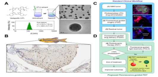
Brain cancer, glioblastoma (GBM), is among the most heterogeneous, treatment resistant, and lethal of all human cancers. The complexity of GBM and the slow progress in effectively treating this disease warrant new combination treatment strategies that leverage imaging tools and technologies earlier in the management of disease. Here, we propose to test an image-guided treatment regimen where we use a new photosensitizer drug and fluorescence imaging to improve photodynamic therapy (PDT) for GBM. An expert team of a cancer biologist (Gottesman), biomedical engineer (Huang), UMD BIOE M.D., Ph.D. student (Quinlan), and a CCR scientist (Robey) will develop a nanoformulation of photosensitizer verteporfin (NanoVP) for fluorescence-guided PDT of GBM in a zebrafish model (Figure 1).
Development of a deep learning algorithm to predict T cell receptor complex structures from sequence, and application to neoantigen-specific T cell receptors
PRINCIPAL INVESTIGATORS:
Brian Pierce, Department of Cell Biology and Molecular Genetics, University of Maryland
Paul Robbins, Center for Cancer Research, National Cancer Institute
PH.D. STUDENT:
Rui Yin, Program in Biological Sciences, University of Maryland
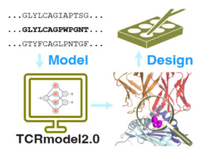
High resolution structures of T cell receptors (TCRs) in complex with their peptide-MHC targets provide critical insights into the mechanistic basis of those key interactions, which are increasingly becoming the focus of clinical and pre-clinical therapeutic studies for cancer. Our team has recently performed initial development of a deep learning-based method to model TCR-peptide-MHC complex structures from sequence, and in this proposed work, we will perform optimization of that method and utilize it to model a set of therapeutically relevant TCR-peptide-MHC interactions that have been identified by the NCI. These models will be assessed for key interface features, including the engagement of the neoantigen mutant residue, and used for structure-based design to validate the models and optimize selected interactions. This algorithm, which will be shared with the community through a web server, will be of high interest to those exploring TCRs for cancer therapy, and these developments can be used to prioritize and optimize therapeutic TCRs in future studies..
Predicting the fate of pre-cancerous lesions observed in long term live animal intravital microscopy
PRINCIPAL INVESTIGATORS:
Wolfgang Losert, Department of Physics and IPST, University of Maryland
Roberto Weigert, Center for Cancer Research, National Cancer Institute
PH.D. STUDENT:
Shuyao Gu, Program in Physics, University of Maryland
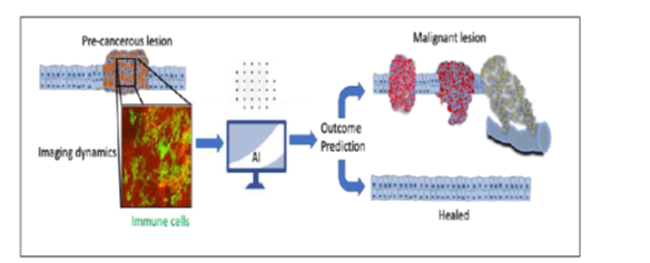
The goal of this project is to analyze the interactions between pre-cancerous lesion and the immune system that Dr. Weigert’s group is now able to image in three dimensions intravitally over long times in a carcinogen model for oral cancer. Based on such data analysis, Dr. Losert will guide the development of AI-based predictive analytics that provides a prognosis of future progression of pre-cancerous lesions into aggressive carcinomas. To this end, the student will acquire and analyze intravital 3D microscopy data during carcinogen-induced tumor development over long times that provide unprecedented insights into the processes underlying cancer progression at a cellular and sub-cellular level. From the measurements, our aim is to characterize how immune cells move and cooperate during natural tumor development.
Mechano-metabolic signatures of metastatic disease
PRINCIPAL INVESTIGATORS:
Giuliano Scarcelli, Fischell Department of Bioengineering, University of Maryland
Kandice Tanner, Center for Cancer Research, National Cancer Institute
PH.D. STUDENT:
Chenchen Handler, Program in Physics, University of Maryland
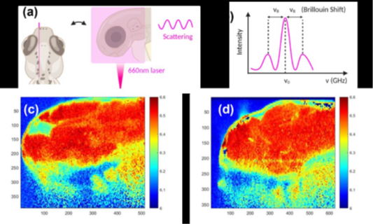
Biological or genetic mutations have been used to identify cancer from normal tissue and to stratify it into molecular sub-types to aid in treatment decisions. In a similar vein, the mechanical property of cells and tissue are considered promising as a potential biomarker since neoplastic and cancer cells progressively show distinct mechanical phenotypes compared to their normal pre-cursors. Recent studies have also shown a critical crosstalk between mechanical properties of cancerous tissues and the metabolic states of tumor cells, widely considered to be an “hallmark of cancer”. Investigation of this promising direction is hampered by our current inability to characterize mechanical phenotypes in tumors in vivo. Specifically, since the interaction between cells and microenvironment is complex and dynamic, traditional mechanical testing methods are not suitable since they are based on contact, they are invasive, and/or limited to the global analysis or the analysis of a few points of a sample. This proposal addresses this important gap thanks to the collaboration between Tanner Lab at NCI and Scarcelli Lab at UMD. Scarcelli Lab has developed Brillouin light microscopy, a technique that allows us to probe cancer cells in native, intact microenvironment and to map their properties within a tissue. Tanner lab has developed a preclinical model of cancer based on zebrafish, which is uniquely compatible with light microscopy, thus enabling assessment of cancer progression over time without perturbation. Thanks to this synergistic expertise, the two labs will investigate the critical link between cancer mechanobiology and metabolism, with the objective to develop predictive biomarkers of metastatic progression based on alteration in tissue mechano-metabolism. The two labs have an established track record of fruitful collaboration; for this proposal, they recruited an engineering graduate student, Chenchen Handler, who has worked in Scarcelli lab since her undergraduate/master studies, has already published a Brillouin microscopy paper as first author, and has collected preliminary data for this proposal (see Fig. 1).
Intracellular dynamics of Argonaute 2 in proliferating and quiescent cells
PRINCIPAL INVESTIGATORS:
Arpita Upadhyaya, Department of Physics, University of Maryland
Joana Vidigal, Center for Cancer Research, National Cancer Institute
PH.D. STUDENT:
Aurelia Moses, Program in Biophysics, University of Maryland
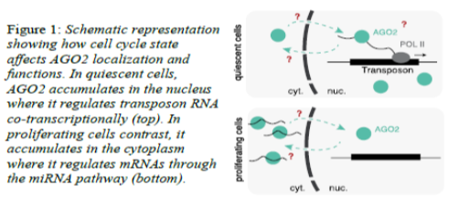
Quiescent cells have temporarily exited the cell cycle. They proliferate in response to injury or stress in order to repair tissues or fight external insults. As a consequence, quiescent cells are essential to the maintenance of organ homeostasis, yet the mechanisms that regulate it remain poorly understood. We have recently found that Argonaute 2 (AGO2), a cytoplasmic component of the miRNA pathway in proliferating cells, translocates to the nucleus when cells enter quiescence. In the nucleus of quiescent cells, AGO2 associates with chromatin of transposable-elements (TE) in an RNA-dependent manner and represses the expression of those loci through its conserved but poorly understood catalytic domain. This proposal aims to understand how changes in cell-cycle state impact AGO2’s sub-cellular localization and consequently its functions as either a cytoplasmic post-transcriptional regulator of mRNAs or a nuclear co-transcriptional regulator of repetitive sequences (Fig 1). This research will combine the expertise of the Vidigal lab (NCI) and the Upadhyaya lab (UMD) and has the potential to uncover fundamental mechanisms underlying the regulation of AGO2 as well as the quiescent cell state, both of which have been intimately connected to tumorigenesis.
Evaluation of low-dimensional image representations in real-time-cryo-EM of cancer structural biology targets
PRINCIPAL INVESTIGATORS:
Wojciech Czaja, Department of Mathematics, University of Maryland
Hans Elmund, National Cancer Institute/CSB Frederick MD
PH.D. STUDENT:
Canran Ji, Program in Mathematics, University of Maryland
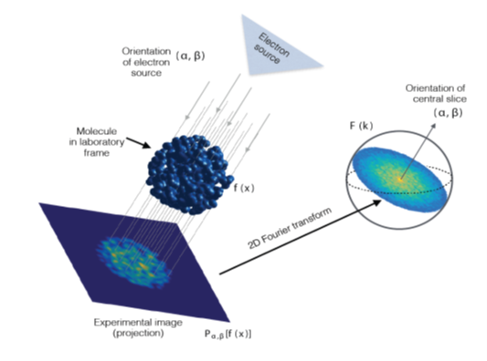
An electron microscope (EM) takes pictures of nanoscale objects, such as viruses that infect our cells or machinelike molecules that catalyze the reactions that are required for our cells to reproduce. We isolate these molecules using genetic and biochemical approaches and flash freeze them in thin films of water-based solutions, which leads to the water molecules creating a glass-like transparent matrix that allows the embedded molecules to be imaged with an EM. The EM takes pictures that are like those that medical doctors obtain using X-rays, representing all the internal structure of the imaged objects. The need for development of computational techniques for determining the relative 3D orientations of the 2D EM images and reconstruct 3D models of the imaged molecules arises, because they are randomly oriented in the ice-layer.
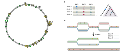
Strain-resolved characterization of cancer microbiome using long-read metagenomic sequencing
PRINCIPAL INVESTIGATORS:
Mihai Pop, Department of Computer Science, University of Maryland
Mikhail Kolmogorov, Center for Cancer Research, National Cancer Institute
PH.D. STUDENT:
Ataberk Donmez, Program in Computer Science, University of Maryland
Cancer is traditionally viewed as a disease of the (human) genome, however there is increasing evidence that microorganisms may play an important role in cancer development, progression, treatment response and diagnostics. The role of microorganisms in cancer development may be understudied in part because of our incomplete understanding of the diversity and function of the human microbiome, hindered by the difficulties in generating and completing bacterial genome sequences. In this proposal, we aim to improve the power of the cancer-microbiome association studies by taking advantage of the complete, strain-resolved bacterial genomes. We will develop a new algorithm for strain-level microbiome reconstruction using long reads and apply it to explore the strain-level evolution and diversity of human microbiome communities and their association with various cancers.
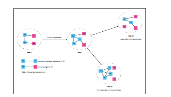
Uncertainty in the Therapeutic Response is Resultant from Intratumoural Heterogeneity
PRINCIPAL INVESTIGATORS:
Wade Winkler, Department of Cell Biology and Molecular Genetics, University of Maryland
S. Cenk Sahinalp, Center for Cancer Research, National Cancer Institute
PH.D. STUDENT:
Chih Hao Wu, Program in Biological Sciences, University of Maryland
Immunotherapy is a relatively new class of treatments for cancer that can be highly successful in some patients, while not generating response in others. We propose to investigate potential modulators of treatment response by analyzing mutated tumour gene sequence features, gene expression regulatory elements, and immune system features that respond to tumour growth. Using established tumour cell lines and mouse models that have had genetic characterization done through previous studies, we will be able to accurately establish the differential effect of these features on immunotherapy response using both established and novel analysis methods.